AFTER EYELID SURGERY
Recovery After Eyelid Surgery
Recovery after eyelid surgery is not assessed by a single day or fixed timetable. Early changes around the eyes, oedema and tissue adaptation are parts of a process that develops over time.
By Mehti Şalvız
Published:
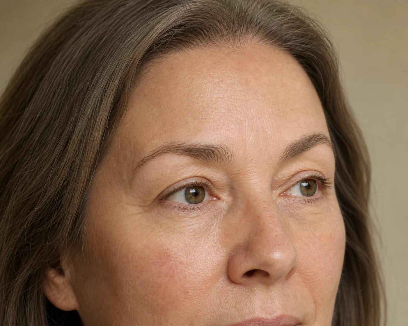
The visible appearance of recovery after eyelid surgery is considered differently according to whether the surgical assessment includes only the upper eyelids, only the lower eyelids or both, and according to the appearance of neighbouring tissues. Although blepharoplasty is a shared term, the upper and lower eyelids are not the same anatomical area; their relationships with surrounding tissues also differ.
Early swelling, oedema, bruising or tissue tenderness may have different distributions across the small neighbouring areas around the eyes. The appearance of the upper eyelid crease and the lower eyelid's transition into the upper cheek are not interpreted through the same feature. Healing tissues may initially make these boundaries appear different from usual.
The following combines general recovery information with care and follow-up information from Assoc. Prof. Mehti Şalvız's eyelid practice. Shared timeframes for upper and lower eyelids do not mean that all surgical details are the same for both.
Why Is Recovery After Eyelid Surgery Different for Everyone?
In the upper eyelid, skin, the eyelid crease, surrounding soft tissues and the visual relationship with the brow are prominent. In the lower eyelid, the contour, under-eye tissues and boundary continuing into the upper cheek form a different anatomical whole. These initial characteristics provide the main context for why early recovery does not look the same in two people.
The area observed when surgery involves only the upper eyelids is not equivalent to the areas in a plan involving only the lower eyelids or both. Including both regions in the same plan does not mean their recovery appearances will progress in parallel; each eyelid is followed in the context of its own tissues and neighbours.
Natural differences between the two sides around the eyes, skin and soft-tissue characteristics and previous procedures accompany assessment. Where swelling, bruising or tenderness seems more prominent is understood alongside this initial appearance and the regions addressed.
Age or another person's experience is not, on its own, a measure of how the eyelids will look. Individual recovery is interpreted through the eyelid assessed, its initial anatomy and the changes observed during follow-up.
What Early Changes May Be Seen Around the Eyes?
Early recovery may involve swelling, oedema, bruising and tissue tenderness around the eyes. Assessment distinguishes the upper eyelid surface, crease, lower eyelid contour and upper cheek boundary; the small areas around the eyes are considered according to their individual characteristics.
The doctor notes that, when canthopexy is added to lower eyelid surgery, the eye corner may appear more lifted early on. Swelling may be somewhat greater when both upper and lower eyelids are treated. These observations do not imply the same course in every patient or a definite time for resolution.
Swelling and oedema
Swelling and oedema may not have the same distribution in the upper and lower eyelids. While the visibility of the crease and upper eyelid area may be affected, the lower eyelid's under-eye contour and transition into the upper cheek may be perceived differently. The right and left sides do not have to look identical at every follow-up.
The location of oedema around the eyes and its effects on neighbouring boundaries are not assessed solely by its amount. Follow-up observes how the relationship between the eyelids' own contours and surrounding tissues changes throughout the process; early fullness or less distinct boundaries do not define the result on their own.
Bruising and tissue tenderness
Bruising or tenderness may not create a uniform appearance throughout the eye area. Thin, mobile eyelid skin, the different anatomy of the upper and lower regions and the scope of the area assessed contribute to where these findings are more noticeable.
The doctor notes that pain may be mild on the first day and that slight eyelid oedema may occur. This observation from his practice is not a guarantee of painless recovery. Report new or increasing pain to your team rather than treating it merely as part of the appearance.
The Early Appearance of the Eye Area
Early on, the upper eyelid crease may be less distinct, the boundary between the lower eyelid and upper cheek may look different from usual, or surrounding contours may appear fuller. Although these areas are close together, they do not represent the same anatomical structure, and changes in appearance are interpreted separately.
Gaze direction, facial expression, lighting and the camera angle may emphasise small contour differences around the eyes. Early assessment is based on observing the relationship between the eyelids and surrounding tissues at subsequent follow-ups, rather than drawing a completed result from one image.
How Are Oedema and Swelling Assessed Over Time?
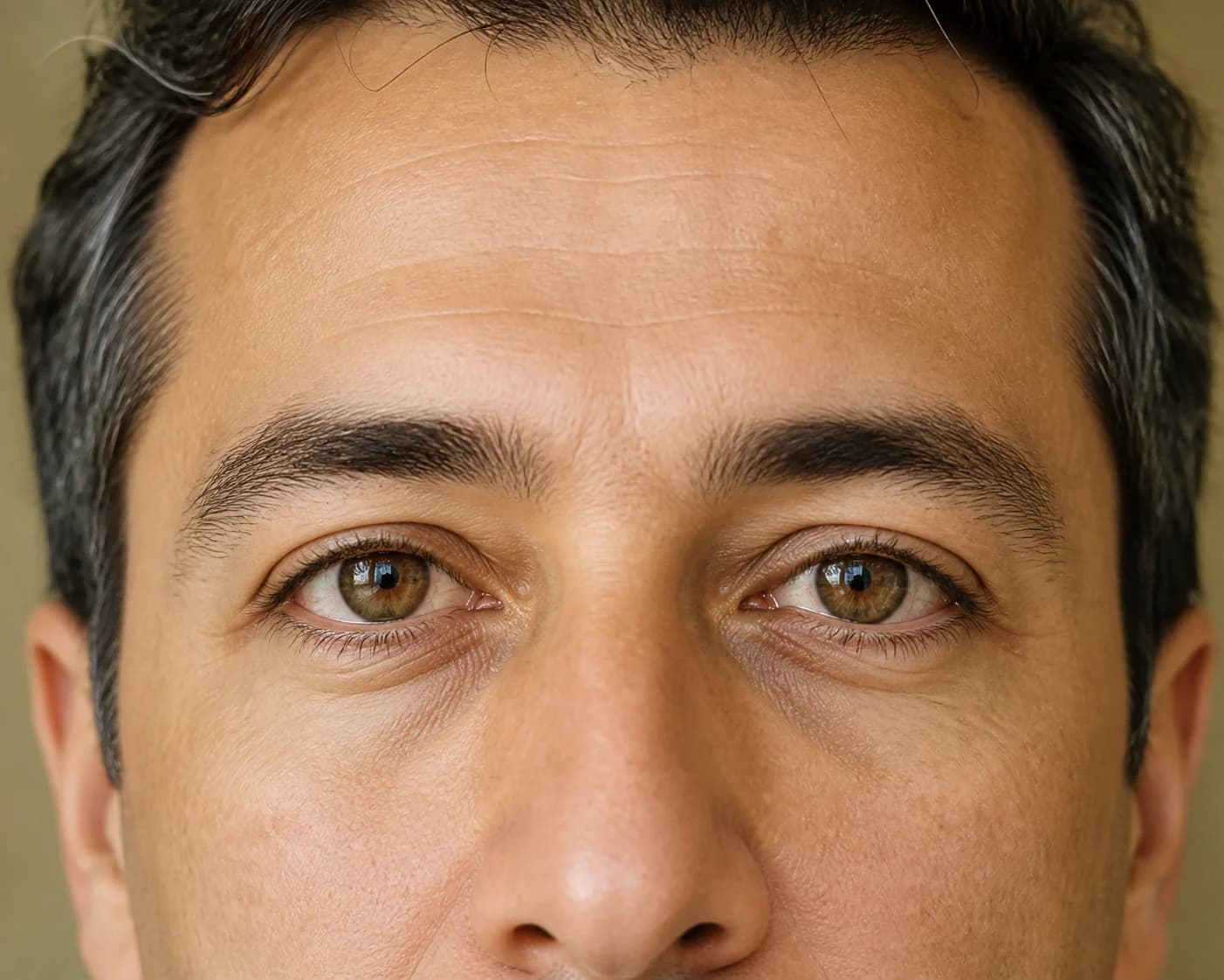
Oedema is not assessed simply as 'present' or 'absent'. The upper eyelid crease and the area beneath the brow, the lower eyelid contour and its transition into the upper cheek are observed within their respective regions. These neighbouring areas are not expected to appear equally defined at the same time.
The impression in a mirror or photograph may vary with lighting, gaze direction and facial expression. At follow-up, the important considerations are comparison with the upper and lower eyelids' initial characteristics and interpretation of their relationships with surrounding tissues, rather than assigning meaning to a single image.
Contact the surgical team without delay if you develop new difficulty closing your eyes, changes in vision, severe pain or unexpected swelling. Do not assume that these findings will resolve on their own.[1]
What Does Tissue Adaptation Around the Eyes Mean?
Tissue adaptation around the eyes describes how healing eyelid tissues form an appearance with neighbouring anatomical areas again. The perception of the upper eyelid crease and area beneath the brow, and the lower eyelid's under-eye contour and boundary extending into the upper cheek, are observed separately within this framework.
The term does not refer only to reduced oedema or tissues suddenly reaching a particular position. As eyelid skin and soft tissues heal, the crease, lower eyelid line and eyelid–cheek transition may appear more defined or softer at different stages. Assessment focuses on changes in these regional relationships.
The general scope of surgical assessment around the eyes is explained on the Eyelid Surgery page. For the separate surgical framework of the upper eyelid, see the Upper Eyelid Surgery page.
The anatomical assessment framework for the lower eyelid and upper cheek transition is available on the Lower Eyelid Surgery page. This lower-eyelid framework avoids assuming the same pattern of appearance as in the upper eyelid; the region assessed depends on the individual surgical scope.
Why Does Returning to Daily Activities Vary Between Individuals?
The doctor's approximate timeframe for returning to general daily life is 5–7 days. This number does not provide separate clearance for exercise, contact lenses, make-up or driving. Vision, eyelid condition and the requirements of the activity are assessed at individual follow-up.
How the person feels contributes to assessment, but the decision is not based solely on that observation or another person's experience. The state of the eye area at that follow-up and the individual recommendation of the doctor overseeing surgical recovery are decisive.
Daily activities do not all have the same requirements; returning to them is considered through individual follow-up rather than a fixed online timetable.
Why Is Follow-up After Surgery Important?
Postoperative follow-up considers not only the external appearance of the upper and lower eyelids but also their visual relationships with each other and surrounding tissues. The upper eyelid crease, lower eyelid contour, regional distribution of oedema and eyelid–cheek boundary are observed according to the surgical scope.
Which change the person noticed, when they noticed it and which appearance they have questions about help guide assessment. This moves beyond simply asking whether swelling is present; the eyelids' individual characteristics and the overall appearance around the eyes are interpreted during the same consultation.
The doctor usually uses non-absorbable sutures and removes them at approximately one week; he does not apply a bandage. Follow-ups are scheduled at one week, one month and six months. Care instructions and the need for additional reviews are arranged according to individual findings.
A change that causes concern should be shared with the doctor overseeing surgical follow-up.
When Are Eyelid Surgery Results Assessed?
Assessment of results is not reduced to whether swelling has decreased. The upper eyelid's appearance formed by skin, crease and brow, and the lower eyelid's contour continuing through under-eye tissues into the upper cheek, are examined within their respective anatomical frameworks. Treating both eyelids at the same time does not mean these relationships will become clearer in the same order.
The doctor uses approximately one month for assessing appearance and approximately six months for assessing the final appearance of scars. Follow-up findings are interpreted alongside these timeframes; they do not guarantee that scars will disappear completely.
The article Differences Between Upper and Lower Eyelid Surgery provides further context for why the upper and lower eyelids require different anatomical assessments. The broader surgical framework covering different regions of the face is explained on the Facial Rejuvenation Surgery page.
The early appearance around the eyes is monitored without assuming that regional healing of the upper and lower eyelids is complete.
Sources
- NHS. Eyelid surgery
Accessed on 6 September 2026. Used for general information about risks and seeking help.
This content provides general information and does not replace individual medical assessment.