Case study / Mehti Şalvız
Endoscopic Deep Plane Facelift: A Case Study
5 min 46 sec · English narration
YouTube ·
In this video, I explain the endoscopic deep plane approach, the patients for whom I prefer it, and images of one of my patients on the second day after surgery and at approximately six months.
The day-two and approximately six-month images in the video show one patient. Healing and outcomes vary between individuals; the appropriate approach is determined through personal examination.
Watch these chapters
Links open the relevant moment on YouTube in a new tab.
- 00:00 — Scope of the approach ↗
Assessment beyond the midface, including the lower face, temples and forehead.
- 01:00 — Incision locations and the deep plane ↗
Incisions within the hair, ligament release and tissue repositioning.
- 02:49 — Endoscopic visualisation ↗
Viewing the surgical area with a camera.
- 03:22 — Day two and approximately six months ↗
Assessment of this patient’s early postoperative and follow-up images.
- 04:27 — Patient selection ↗
How excess skin and laxity influence the choice of method.
How do I assess the technique and this case?
The following text is an English translation of the existing Turkish adaptation of Assoc. Prof. Mehti Şalvız’s video explanation, rather than a verbatim transcript. The assessments relate to the case shown in the video.
More than a midface-only procedure
In this video, I explain the endoscopic deep plane facelift approach through one of my patients. The procedure I am describing is not simply an endoscopic midface lift. My assessment also includes the lower face, midface, temples and, where needed, the forehead.
I show the boundaries of the area we work in on an anatomical drawing. I consider the region extending down to the jawline together with the upper face; the approach to the forehead is planned according to the patient’s needs.
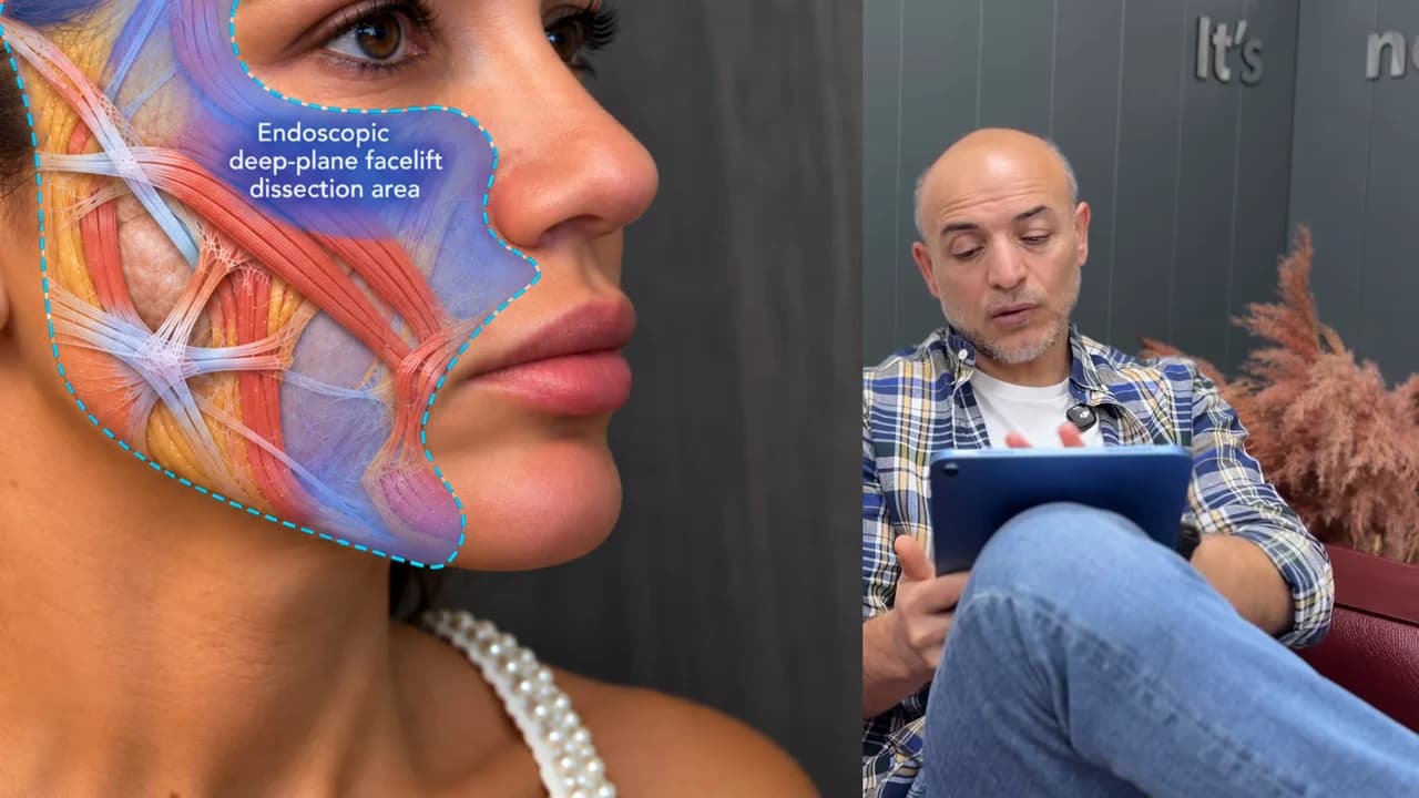
Incisions within the hair and deeper tissues
In the endoscopic approach, I use incisions within the hair in the temple region. Additional incisions for the forehead can be planned when needed. The approach I describe in this video does not use the incision in front of the ear found in the traditional method.
By releasing the ligaments that hold the tissues, I can mobilise the deeper structures. As in the traditional deep plane approach, our working plane is beneath the SMAS layer. Here, I aim to reposition the tissues in a more vertical direction.
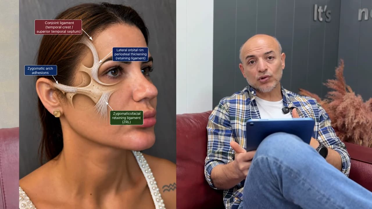
Viewing the surgical area with an endoscope
The endoscope lets me see the area I am working in with a camera. I use this view to follow the tissues and the ligaments I release. The drawings in the video explain the anatomical structures beneath the changes we see at the surface.
In my assessment, I consider the folds extending from the nose to the corners of the mouth, the lower eyelid area, brow position and hooding above the upper eyelid together. I do not assess these as independent areas.
This patient’s second postoperative day
In the video, I show images of my patient on the second day after surgery. These show mild swelling and some bruising around the eyes. I assess this early appearance as one stage in this patient’s recovery.
I do not treat the day-two image and later follow-up images as if they were taken at the same time. In the video, I show each within its own recovery period.
Assessment at approximately six months
In the later comparison, I look at images taken at approximately six months. I assess the folds beside the nose, the lines extending down from the mouth corners, brow position and the eye area alongside the preoperative images.
In explaining this case, I consider the appearance of the lower face as well as the midface and eye area. The comparison in the video is based on the same patient’s initial and follow-up images.
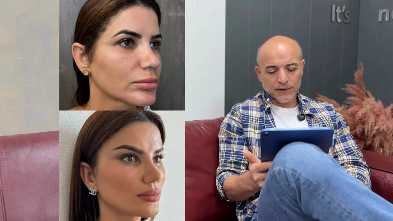
Excess skin is a key factor in choosing the method
I do not recommend this approach in the same way for every patient. Regardless of age, I look at the amount of excess skin, tissue laxity and sagging along the jawline. In the video, I particularly emphasise the importance of excess skin in this decision.
When there is little excess skin, I can consider an endoscopic approach. With more substantial excess skin, a traditional deep plane approach requiring an incision in front of the ear may be considered to remove it. I decide which technique is appropriate by assessing the patient’s own anatomy.