AFTER DEEP PLANE FACELIFT
Recovery after deep plane facelift surgery
Recovery after deep plane facelift surgery is not a fixed timetable predicted from the technique name alone. Early tissue changes, swelling, surgical scope and facial anatomy are assessed together as recovery is followed over time.
By Mehti Şalvız
Published:

Recovery after deep plane facelift cannot be treated as a fixed timetable that progresses identically for everyone based on the method’s name. Deep plane describes an anatomical approach in facelift surgery; postoperative appearance is shaped by facial anatomy, surgical scope, the areas addressed and individual tissue response.
Swelling, oedema, bruising or tissue tenderness may occur early on. Their location and effect on contour can vary between individuals. The midface, lower face, jawline and, when involved, upper neck need not show the same appearance at the same time. Early facial contour is therefore not interpreted as the completed result.
A broader explanation of recovery topics shared after facelift surgery is available in the Recovery after facelift surgery article.
Why can recovery after deep plane vary between individuals?
Recovery appearance requires consideration of both starting anatomy and the tissues’ response to surgery. Skin thickness, soft tissue distribution, facial contour and healing characteristics can differ. None of these alone predicts exactly how recovery will progress; their significance emerges within the full clinical assessment.
Surgical scope also matters. One plan may focus on the midface and lower face, while another includes the relationship between the jawline and upper neck. Sharing an approach name does not mean every region and tissue is addressed to the same extent. Visible swelling, tenderness and contour changes therefore cannot be compared against a single example.
The deep plane approach is a particular anatomical context within the broader assessment of Facelift Surgery . Associated neck assessment, previous procedures, general health and individual tissue responses observed during follow-up are other considerations. These variables provide the context for the anatomical areas in which early appearance is monitored.
What changes may occur early on?
The early period is when healing responses begin in the surgical areas and appearance can vary. Swelling, oedema, bruising and tenderness are followed in the context of the areas addressed, tissue characteristics and individual surgical plan.
Swelling and oedema
Swelling and oedema can be visible aspects of the early tissue response to surgery. The midface, cheeks, lower face and jawline need not be affected equally. A fuller-looking region or less defined boundaries do not mean the whole face is healing identically or that the result can already be judged.
The amount and distribution of swelling cannot be attributed simply to “deep plane”. Facial anatomy, the areas assessed and individual tissue response may jointly influence appearance. The two sides may not look entirely alike at the same stage; regional distribution is followed in relation to the midface, lower face and jawline within the surgical scope.
Bruising and tissue tenderness
Bruising and tenderness may occur early on; their visibility and distribution vary between individuals. Skin characteristics, the operated areas and tissue response can influence how they appear. More or less pronounced findings in another person do not provide a measure for individual recovery.
Assigning tenderness a numerical scale or standard timetable is outside the scope of this general information. No medication or home-care protocol is provided here. Changes observed by the individual are assessed in their own recovery context by the doctor who knows the surgical plan.
Early appearance of facial contour
Early facial contour can be affected by swelling and healing responses. The midface, lower face, jawline and upper neck transition may not all have a settled appearance at once. Fuller areas or less distinct transitions may reflect the tissue appearance at that stage.
Assessment of early contour considers how transitions between facial regions change during follow-up. A single photograph does not fully show tissue quality, swelling distribution or these transitions at different stages. Relationships between the midface, lower face and jawline are considered together.
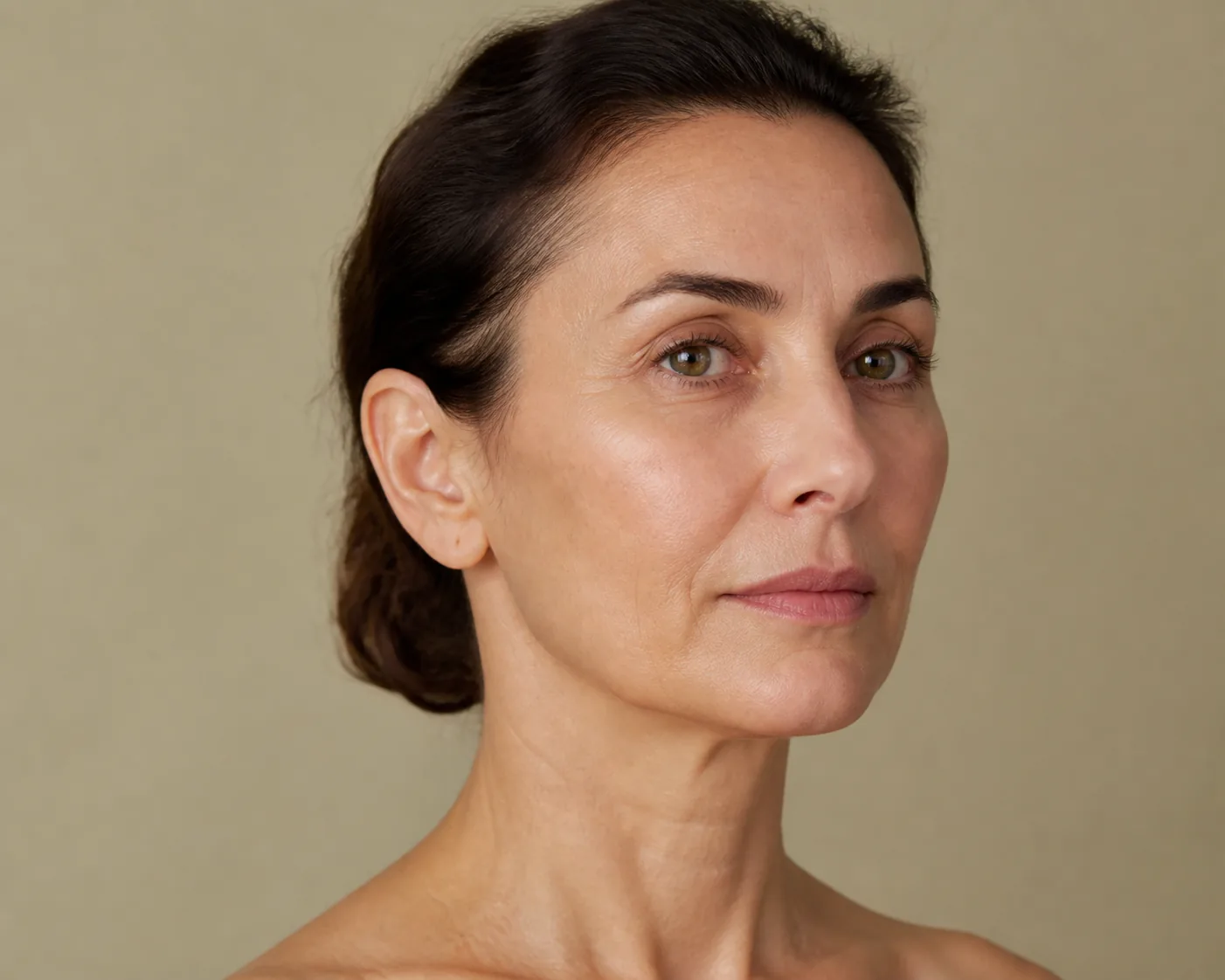
How is swelling after deep plane assessed?
The name deep plane provides no fixed measure of how much swelling will occur, where it will be most apparent or how long it will last. Oedema is a change that can occur during healing after surgery; its appearance is considered alongside anatomy, the areas addressed and individual healing response. Even within the same approach, its distribution in the midface, lower face or jawline may differ.
Swelling’s effect on contour does not stay the same at every moment. Lighting, facial angle and daily conditions can change its perceived appearance, but these observations are not criteria independent of clinical assessment. Change in one region is related to neighbouring areas and the surgical scope.
Changes in distribution across the midface, lower face and jawline are followed in relation to the individual surgical plan.
Why can surgical scope affect the appearance of recovery?
Although “deep plane” describes a particular anatomical approach, it does not define a standard procedure list involving the same regions to the same extent in everyone. The plan is shaped by anatomy, distribution of changes and which areas need assessment together. A midface-focused assessment differs in scope from one also addressing the lower face, jawline or upper neck.
Recovery appearance cannot be considered independently of this scope. The number and relationship of operated areas and individual tissue characteristics can contribute to perceived swelling, tenderness and contour. This is not a classification implying that a particular scope automatically means easy or difficult recovery.
The anatomical assessment and surgical planning framework of the approach is explained on the Endoscopic Deep Plane Facelift page.
Does endoscopic access mean faster recovery?
No; endoscopic use alone does not mean faster recovery. Endoscopic refers to visualisation and access in the surgical area. Deep plane concerns an anatomical plane in facelift surgery. The terms do not describe the same thing and have different roles in assessing recovery.
Recovery appearance cannot be reduced to the access approach. Surgical scope, facial areas addressed, anatomy, skin and soft tissue characteristics and individual healing response are considered together. Endoscopic use therefore cannot promise less swelling, more limited bruising or easier follow-up.
How these concepts differ and the framework for assessing facelift approaches are discussed in the Deep Plane and Other Facelift Approaches: The Differences article.
Recovery in the doctor’s endoscopic practice
The following information from Assoc. Prof. Mehti Şalvız concerns his endoscopic deep plane practice, which mainly addresses the upper and midface without substantial skin removal. It should not be generalised to every deep plane plan involving pronounced neck and jawline sagging or more extensive skin removal.
This procedure takes approximately two hours under general anaesthesia, with discharge planned for the following day. A bandage is used for the first two days. Mild pain and nausea may occur on the first day, with swelling and bruising around the eyes early on.
The doctor anticipates approximately one week before returning to general daily life. Sport, makeup and driving are assessed separately. The follow-up visit he specifies is on the third day after surgery. Other details of follow-up should be clarified with the team.
In this practice, the doctor uses approximately one month for the appearance to become clearer and approximately six months for assessment of the final appearance. These intervals do not promise a definite result or faster recovery for everyone.
How are tissue adaptation and changes in facial contour assessed?
Tissue adaptation is a general expression describing the changing appearance of operated areas as they heal over time. It is not limited to changes in swelling. Healing of skin and soft tissues, changes in the perception of transitions between facial regions and evolving contour during follow-up are considered together.
Tissues should not be thought of as mechanically “settling into place” all at once. The midface, cheeks, lower face, jawline and upper neck may not show the same appearance simultaneously. A different course in one area does not alone indicate a positive or negative result. Appearance is assessed alongside starting anatomy and surgical scope.
A broader framework for the relationship between facial regions within one plan is available on the Facial Rejuvenation Surgery page.
Why is follow-up important?
Follow-up helps avoid interpreting recovery only from external appearance or a person’s impression at one moment. The distribution of swelling, tissue tenderness, contour changes and the overall appearance of operated regions can be assessed within the individual plan.
The person’s observations and questions are also part of follow-up. Appearance in the midface, lower face and jawline is compared with the areas included in the plan and previous assessments. Where swelling appears more noticeable is followed within the same anatomical framework. Questions can then be considered in relation to the observed area and individual surgical scope.
Decisions about follow-up, medicines, care and returning to activities depend on the surgery performed. Contact the surgical team without waiting for a routine visit if unexpected swelling, fever, severe pain, skin changes or trauma develop later.
When are results after deep plane assessed?
Results are assessed through the transition from the midface and cheeks to the lower face, definition of the jawline and, when relevant, its relationship with the upper neck. The effects of swelling and tenderness on these contours are considered regionally and alongside the overall appearance during follow-up.
These regions may not change to the same degree. Follow-up compares the overall contour relationship between the midface, lower face, jawline and upper neck rather than one area alone. Different observations help show how the contour formed by these transitions is perceived. Starting anatomy, surgical scope and tissue appearance provide the context.
Long-term durability is a separate subject for assessment.
This content provides general information and does not replace individual medical assessment.